Shoulder arthritis
What is it?
Arthritis is wear and tear of the joint. Osteoarthritis is a slow process that develops over a number of years. With this type of arthritis the joint cartilage gradually becomes thin and roughened. The bone underneath thickens and the bone at the edge grows outwards causing bony spurs (osteophytes). The joint may swell as the body makes extra fluid to lubricate the joint. The joint may get deformed and there will be gradual loss of movement.
What is its cause?
The most common cause of osteoarthritis in the body is wear and tear of the joint due
to ageing. This is called ‘primary osteoarthritis’. Fractures or instability of the shoulder can lead to arthritis later in life. This is termed as ‘secondary arthritis’.
In the shoulder a specific of arthritis known as Rotator cuff arthropathy can develop. It occurs in patients with large, long-standing cuff tears which subsequently develop in to arthritis between the humeral head and acromion. The shoulder is a ball (humeral head) and socket (glenoid) joint. It is raised, lowered or rotated by a group of four muscles (supraspinatus, infraspinatus, subscapularis and teres minor), which are called the ‘rotator cuff muscles’. They also help to stabilize the shoulder joint i.e. they try to keep the ball at the centre of the socket. In the presence of a cuff tear, the ball of the shoulder (Humeral head) will become unstable and move upwards towards the acromion(Shown in Figure below). With time this may cause arthritis between the humeral head and the acromion (tip of the shoulder blade).
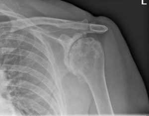
What are the symptoms and how is the condition diagnosed?
The symptoms are of gradually increasing pain and stiffness. With time the symptoms increase and cause significant functional disability. The patient may complain of grinding or crunching within the shoulder. In patients with rheumatoid arthritis the shoulder can be damaged at any age. Patients with rheumatoid disease may have involvement of the other joints in the limb.
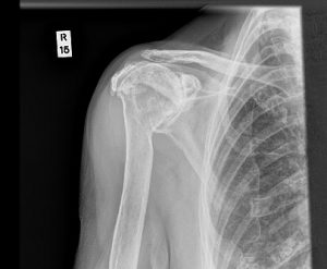
What investigations will be needed?
An x-ray of the shoulder is needed. It will confirm the extent and type of arthritis.
A CT scan may be advised to assess loss of bone due to arthritis, to decide on the right type of replacement. A MR scan may be advised to assess the integrity of the rotator cuff.
What is the treatment?
1. Initial management consists of painkillers, anti-inflammatory medication and activity modification. It is aimed at easing the pain and regaining motion.
2. If the above measures fail then surgery is considered. The type of surgery that will depend on the nature and longevity of the symptoms, type and extent of arthritis, age and functional demands of the patient.
Arthroscopic (keyhole) surgery: A relatively small operation during which a Telescope is passed into the shoulder through one small incision (½ cm).
Additional 1-2 small stab incisions are used to pass instruments, to allow for removal of loose bodies, inflammatory tissue and debride any loose cartilage.
Such arthroscopic debridement will provide temporary relief.
Shoulder replacement: The eventual solution to an arthritic shoulder is to replace it. Different types of shoulder replacements can be used, the decision of which isbased on age, functional demands of the patient, extent of arthritis, any bony loss and presence of a rotator cuff tear.
Surface replacement: In this type of shoulder replacement, just the humeral head is replaced. The advantage of this procedure is that it preserves the maximum amount of bone and minimal soft tissue dissection is needed. If the replacement fails in the long-term (all replacements will eventually) revision surgery to change the prosthesis is easier.
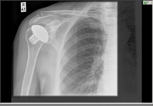
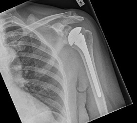
are replaced.
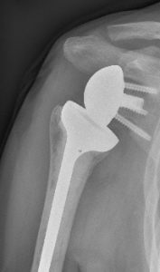
Reverse geometry total shoulder replacement: This procedure in which both the ball and the socket are replaced and reversed, is practiced for rotator cuff
arthropathy. In the presence of a cuff tear, the deltoid is the only functioning muscle. Reversing the normal shoulder medializes the centre of rotation of the joint. This maximises the lever arm of the deltoid muscle and helps achieve a better range of motion of the shoulder.
What happens if it is not treated?
It is possible that with time pain may lessen, but motion between the humeral head and glenoid will be lost. However most patients will find that their symptoms will increase with time and they will need further treatment
What is the success of surgical treatment?
Each of the surgical treatments is successful if they have been advised at an appropriate stage of the disease. More than 90% patients will achieve a significant improvement in their pain after these procedures. Success rates for regaining the movement will vary and you may want to discuss this further with your Shoulder Specialist. The strength in your arm will take longer to improve, and will be dependent on the amount of pain and stiffness you had prior to the surgery.
What are the complications of surgical treatment?
1. The surgical scar may remain unattractive. An area of numbness can occur around the scar but this does not cause any problems.
2. Infection of the wound is possible but usually can be successfully treated with antibiotics. Very rarely infection may spread into the joint replacement.
3. Injury to the nerves (axillary) can occur. This may lead to altered sensations and shoulder / limb weakness.
4. The shoulder replacement may dislocate.
5. In the long term, the shoulder joint replacement may fail. In this case, the replacement will need to be revised. It is expected that nearly 85-90% of shoulder replacements will still be successful after 10 years.
6. The replacement may fail to relieve the pain totally.
7. Shoulder stiffness
8. Fracture of the glenoid or humerus
9. Any surgical intervention has the risk of developing complications / setbacks which are unpredicted. These complications may have the potential to leave the patient worse than before surgery.
Is there anything I can do to improve the outcome?
• Keep the wounds dry and clean until they have healed.
• After the operation the therapist will see you to start movements of the shoulder. It is important to carry out the prescribed exercises regularly, both during the physiotherapy sessions and at home. It will help to keep the pain levels down with analgesics so as to keep your shoulder, elbow and hand fingers moving.
• It is advised against wearing rings on the operated side for 4 to 6 weeks after surgery.
When can I do various activities?
Return to work depends on many factors including the nature of the job and hand dominance. Following shoulder replacement you will be to return to a desk job within 4-6 weeks of the operation and perform reasonable tasks with the limb by that time.
Manual work is not generally recommended after a shoulder replacement.
Driving should be possible within 1 or 2 weeks of keyhole surgery, and within 4 to 6 weeks of shoulder replacements. Before driving, do check that you can manage all controls and start with short journeys.

